PET-MRI scanner opens new frontier in medical imaging
Harry Wood reports on the potential of Philips' new scanner which has recently been inaugurated at the University Hospitals of Geneva.
28 May 2010
The first combined PET-MRI scanner in Europe, and only the second in the world, has been installed at The University Hospitals of Geneva (HUG). The revolutionary device was officially inaugurated at a symposium at HUG at the end of April.
The PET-MRI scanner, which has been developed by Philips, combines magnetic resonance imaging (MRI or IRM in French) and positron emission tomography (PET) into one unit, allowing accurate combination of the two imaging modalities and giving new possibilities for medical imaging.
Currently when both these types of images are needed they are taken separately, often on two different days, making it difficult to align the imaging accurately and combine the properties of each. Combining both units gives greater comfort to patients and makes it simpler and faster for the radiologists to combine the images.
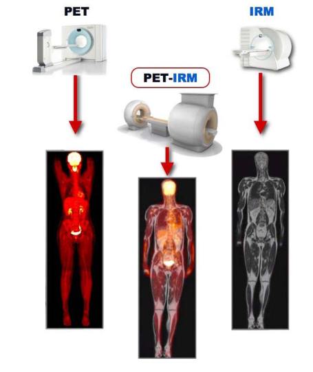
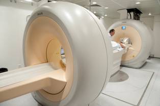
The PET-MRI (IRM in French) scanner combines
the imaging of both modalities in one unit enabling accurate
combination of both images. Source: HUG
Positron Emission Tomography (PET) scanning uses a radioactive tracer that is injected into the blood to send it to the body tissue of interest. The tracer binds to tissue or organs and enables the scanner to produce 3-D images of functional processes in the body, highlighting abnormalities that indicate disease. It is commonly used to monitor certain types of cancer and also for brain activity, eg for detecting Alzheimer's disease. Magnetic Resonance Imaging (MRI) uses a strong magnetic field to excite hydrogen atoms in water in the body. This picks up detailed imaging of the anatomical structure of the body.
The combined techniques will enable specialists to better understand complex diseases such as head and neck cancer, breast cancer, prostate cancer and to monitor the effects of treatments.
Currently the only other Philips PET-MRI scanner is at Mount Sinai Hospital in New York. Both will be used in routine clinical examinations and research projects to test and demonstrate the value of the combined imaging in areas such as neurology, cardiology and infectious and inflammatory diseases.
Combining PET and MRI is not a new idea. The first design was published in 1997 and there have been various developments to try to overcome the interference from the MR magnets on the PET scintillation detectors and integrate the detectors into one unit. Recently photodiodes that are insensitive to magnetic fields have been used as PET detectors. However, previous prototypes have only been built for animal use or just for brain scans. This is the first combined scanner that can do whole-body scans, although the two scanners are physically separated.
.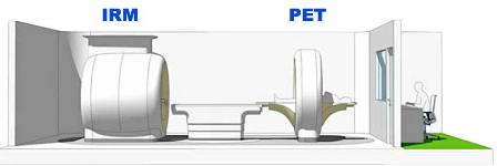
The Philips PET-MRI scanner layout, showing the
separation of the two scanner types by a revolving bed. This layout
prevents interference of the PET imaging by the strong magnetic
field of the MRI scanner and the bed enables easy transfer of the
patient from one scanner to the other and accurate positioning of
the patient in each scanner. Source: HUG.
Professor Osman Ratib, Chair of the Department of Medical Imaging
and Information Sciences at the University Hospitals of Geneva said,
"This new device is combining for the first time two whole-body
scanners that were traditionally impossible to put together. It was
traditionally impossible to put them together because the magnetic
field of the MRI prevents a PET scanner from functioning properly
and creates major artifacts [in the images] that were considered
impossible to overcome for a long time.
"Thanks to this new technology Philips Healthcare has developed, they succeeded to put those two devices into one room separated by one bed where the patient can go from one scanner to another and obtain images that perfectly align.
"MRI images provide anatomy and tissue characterization combined with metabolic imaging obtained from PET, so we can see function and metabolism of the tissue. And thanks to new molecular imaging and molecular tracers that we inject into the patient, we are able nowadays to see and follow some tissue characterization and tissue activities very precisely thanks to hybrid imaging."
"In our collaboration agreement with Philips Healthcare, we have agreed to evaluate the impact and the added value of these hybrid imaging techniques especially in some very specific domains of oncology and cancer follow-ups. We have chosen three domains where we think it may have a major improvement or impact, one of them being for head and neck cancers.
"Patients get very aggressive surgical treatments and it is very difficult to identify recurrence or changes in cancer in those modified anatomies. We believe that adding PET and MRI together in one single superimposed image will be a major improvement compared to what we do today where the two images are being acquired at different times and are sometimes very difficult to overlap or combine.
"The other domain we have selected is for prostrate cancer where it was very important to be able to detect early on recurrence of cancer. We also have the same difficulty combining MRI imaging with metabolic imaging of PET today so we think having those two images superimposed will really improve the quality and the performance of our diagnosis in detecting early recurrence of those cancers.
"The third domain that we selected is for breast cancer where MRI is becoming a major player in detection and follow-up of those cancers. With improvement in technology, we can now have more specific analysis of those cancers especially in very complex cases. We believe that adding the PET images in a perfectly aligned way to the MRI images will add the metabolic dimension to investigations which will allow more precise and accurate detection and diagnosis and follow-up on the effects and efficacy of some of the treatments."
Development
The scanner was developed in just over a year by Philips with the Translational and Molecular Imaging Institute, Department of Radiology, Mount Sinai School of Medicine, New York. There, Dr Zahi Fayad and his team have been studying applications in cardiovascular imaging. They found that the quality of the MR-PET images allows a better visualization and quantification of the vascular beds (mainly aorta and carotids). Also, the use of MRI-PET scanners instead of PET/CT (the current popular combined modality scanner), reduces the extra radiation dose to the patient and gives higher soft tissue contrast, allowing better visualization and understanding of the patient's underlying disease.
Further PET-MRI scanners will be installed in Dresden, Madrid and Sao Paulo over the next year. The five sites will be studying the scanner's application in breast, prostate, head and neck cancers, and cardiology. Philips is looking for sites that could study infectious diseases and diabetic foot using the scanner.
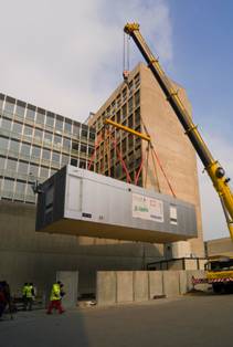
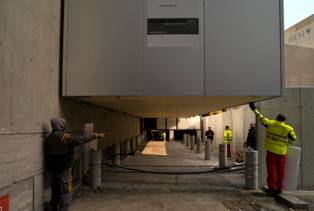
Scanner suite installation
Philips has not just created a novel imaging system, it has also designed a novel delivery system for the device — a complete scanning suite in a container. The container is delivered by truck and dropped into place by crane. The container was built and furnished by Lausanne company X-ion, and at 48 tonnes requires specialist transport and a heavy-lift crane on site to put it in place on ready-prepared foundations and with power, water and drainage ready to connect.
 |
 |
 |
|
| The imaging suite being transported and lifted into position. Source: HUG. | |
Inside the scanner suite. Source: HUG.
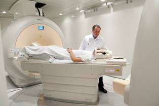
Dr Usman Ratib with his new 'baby' installed
and ready to use! Source: HUG.
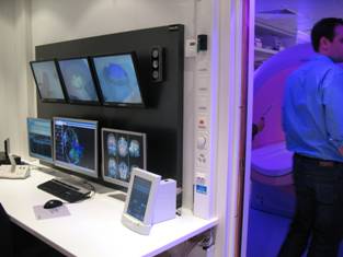
The scanner control room.
© Harry Wood, The Birchley Hall Press.